Cell-cell junctions in the pathogenesis of arrhythmogenic cardiomyopathy
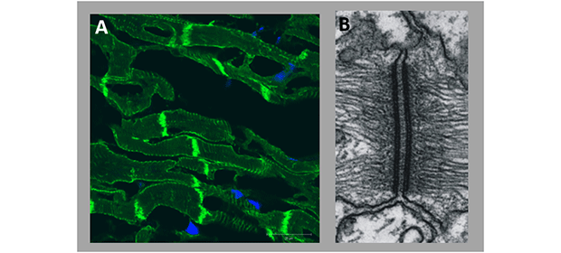
Dominant mutations in genes encoding cellular junction proteins of the cardiac desmosome account for a significant proportion of ACM cases. However, the sequence of cellular and molecular events leading to arrhythmias and heart failure in patients with mutations in desmosomal proteins is poorly understood. Cardiac desmosomes are cellular adhesion structures located at the intercalated discs (Fig.1) that provide the mechanical attachment between cells and consist of members of at least three protein families: desmosomal cadherins including desmoglein-2 and desmocollin-2 (DSG2, DSC2), armadillo proteins such as plakophilin-2 (PKP2) and plakoglobin (JUP) and the intermediate linker protein desmoplakin (DSP).
Recent studies on different mouse models lacking or overexpressing desmosomal components have shown that cardiomyocyte adhesion mediated by desmosomal proteins is a sensitive biological process. For example, we have shown in a mouse model overexpressing DSC2 (Brodehl et al. PLoS One 2017 Mar 24) that changes of the homeostasis between desmosomal cadherins induce a cascade of different cell-cell interactions mediated by a complex network of various extracellular signaling molecules which leads at the end to profound cardiac remodeling and heart failure in mice. However, important questions addressing signaling cascades initiating the remodeling process as well as the selection of target molecules for specific therapies remain open and are our current research Focus.


